What is ACL Reconstruction?
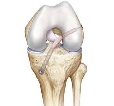
ACL reconstruction is the reconstruction of the anterior cruciate ligament using tissue from elsewhere in the body or from a cadaver. The most common tissue used as grafts for the ACL reconstruction are hamstring tendons, the middle third of the patellar tendon (tissue connecting your knee cap to your tibia), or the quadriceps tendon.
Choice of Graft
The two most common graft choices are the hamstring graft and the patellar tendon graft. Each has advantages and disadvantages and each has excellent long term results.
The hamstrings are used most commonly in British Columbia. Hamstring strength remains excellent with strength deficits in the operative leg varying between 3 and 15% one year after surgery. The major loss of hamstring power is in the last few degrees of flexion and the majority of people do not recognize any deficit in strength at all. Some athletes in certain sports, such as elite sprinters, may be better suited to another graft choice but for most people an ACL reconstruction using hamstring tendons offers an excellent option.
Patellar tendon grafts are also an excellent graft choice and have been used successfully for athletes at the highest level for many years. Patients who receive this graft obtain excellent knee stability. A small piece of bone is taken from the patella (knee cap) and tibia (shin bone) with the patellar tendon in between. Incisions are slightly less cosmetic with this operation. This graft is stiffer and re-ruptures at a slightly lower rate when compared to a hamstring graft. However, there is a higher rate of knee cap pain, and difficulty kneeling due to pain after this procedure. There is also a small but present danger of suffering a fracture to the patella postoperatively.
Many long term comparison studies have been presented over the years comparing these two graft options and most people do well with either type of reconstruction.
Surgical Technique
The reconstruction is performed using an arthroscope which allows for smaller incisions, better visualization and less trauma to the knee.
The graft is harvested through a small incision over the knee (2-3 cm for hamstrings, 5-8 cm for patellar tendon). It is then prepared by your surgeon for insertion into the knee.
Three or four poke holes (3-4 mm) are made in order to place the camera and surgical instruments into the knee. An inspection is made of all parts of the knee to assess for injuries to cartilage, menisci or other ligaments. If damage is found your surgeon with treat it prior to reconstructing the ACL.
The knee is prepared for insertion of the graft by debriding the remnants of the ACL from its insertion. Two tunnels are then drilled in bone, one in the femur and one in the tibia, in order to accept the graft. The tunnels are placed in the ‘footprint’ of the natural ACL so that function is returned optimally.
The graft is then shuttled into the knee and secured using a variety of instruments. Stability is checked at the end of the case before the patient is woken up and the wounds are closed.

Post Operative
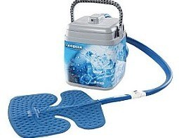
ACL reconstruction is generally a day-care procedure, meaning the patient does not spend a night in hospital. Crutches are necessary for 1-2 weeks and by 6 weeks most people can walk with only a minimal limp. Icing should be performed several times per day for 20-30 minutes or a Cryocuff should be used if it is available. The patient will be provided with a physiotherapy protocol and supervised physiotherapy MUST be started within the first 5-10 days after surgery. Failure to follow the physiotherapy protocol can result in knee stiffness, pain and early failure of the graft. Physiotherapy will be required 1-3 times per week for up to 6 months after surgery.
When first put into the knee, the graft itself has no blood supply. Over a period of time, however, it gains a new blood supply, comes back to life and strengthens. Full graft incorporation takes up to two years to complete. As the body is turning the graft into a new ligament, it actually weakens the graft. Between 3 and 6 months the graft is at its weakest just as the patient is starting to feel well enough to think about going back to sports. The surgeon and physiotherapist will guide the patient through a return to activities in a manner that protects the graft as it heals. Returning to cutting, twisting, pivoting activities should not be attempted until 9-12 months postoperative.

Return to Work: Students and those with desk jobs can generally return 7-10 days after surgery provided that the amount of time spent on their feet is limited.
Patients with jobs requiring significant standing or walking may require up to 6 weeks off. Heavy laboring jobs or jobs where accidental twisting of the knee may occur require 4-6 months before a return is possible.
Risks or Complications
Overall the number of people who have problems following ACL reconstruction is small. Nevertheless, problems do occur and these need some consideration. Below is a list of some of the common problems following surgery but it is not a comprehensive list of all possible complications.
Bruising in the immediate post operative period is not uncommon. Obviously everybody has some bruising, but occasionally, it is such that the knee becomes swollen and sore and the normal mild discoloration that extends to the foot becomes very obvious. This gives discomfort particularly when standing up and may last two weeks. To a degree this can be avoided by ice and rest in the immediate postoperative period.
DVT (deep vein thrombosis) also occur but are uncommon (~1%). These represent clots in the deep veins of the leg, usually the calf. They probably occur at the time of surgery and then get slowly bigger over several days. Because of this they may not be felt in the first few days. If noticeable, it is usually as an ache in the calf at the back of the leg. If this is thought to be occurring, then an ultrasound scan can be used to detect it and appropriate treatment organized.
The concern of having clots in the vein is always that they may spread to the lungs (pulmonary embolism or PE). This is a rare event but does represent the one major and serious complication of this and other lower limb surgery. In the majority of cases, like DVT’s themselves, it is treatable by thinning of the blood. This prevents new clot from forming and allows the body to slowly dissolve the clot that is present.
Infection is uncommon and occurs in about 1 in every 300 cases. Almost all such infections can be treated without loss or failure of the graft. Nevertheless, the graft is threatened by this problem which requires prompt treatment, including arthroscopic washout of the knee and antibiotics. Early diagnosis is very important to avoid damage to the rest of the knee joint.
Knee stiffness can occur after ACL surgery. For the most part it can be avoided with early physiotherapy and close follow-up. Pain and swelling control is important to allow for early range of motion. Cryocuff use (or bags of ice) early postoperatively and early physiotherapy are important to begin early motion and avoid stiffness.
Graft failure re-rupture can and does occur. No graft is as strong as a normal ligament and hence a big enough injury can cause damage to it. The risk of re-rupture varies depending on the patient’s activity profile and age among other things. At two years, the risk of ACL graft rupture is approximately 3-5% depending on a number of factors and in fact the risk of rupturing the contralateral (opposite side) ACL during that time is approximately the same! Physiotherapy is important to train muscles to protect the knee during jumping, landing, turning and twisting in order to minimize the risk of re-rupture.
Patello-femoral pain or ache under the kneecap (patella) is common once activity has begun. This is mostly due to the muscles being wasted and weak and therefore responds well to exercise, particularly of the quadriceps muscle. Physiotherapy at this stage is the treatment for pain at the front of the knee. Patello-femoral pain can also occur from damage to the articular lining of the patella itself. This occurs in some patients either from the injury or from wear and tear over time. Again physiotherapy generally improves the symptoms but with damage under the patella they may not entirely dissipate. This type of pain is slightly more common after a patellar tendon graft.
Patellar tendonitis or ache from the remaining patella tendon, is not uncommon at some stage during recovery when using a patellar tendon graft. Fortunately, this tends to be transient and tends to settle over a 1-2 month period. In most people this occurs when running is commenced and it represents stress on the remaining smaller patella tendon. This stress then stimulates the tendon to get bigger and stronger (hypertrophy) until it is able to cope. With time the tendon usually settles down and stops aching when used.
Ultimately most knees settle down over a 12 month period of time and by that stage most people are no longer conscious of their knee.
